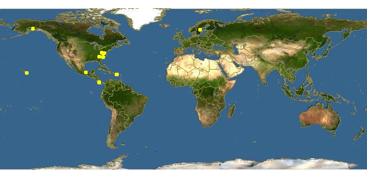
Click on map for details about points.
|
|
Overview | |
Cultivated on substrates such as 0.1L-P, 0.1L-P (pH 6.0), and hay infusion agars in association with Escherichia coli or Escherichia coli B/r at 22-25 C. Sorocarps large and variable, mostly solitary, usually semi-erect or inclined under overhead light and prostrate in unidirectional lateral light, strongly phototropic, 4-10 mm or more high when semi-erect and up to 5-7 cm or more in length when primarily procumbent. Sorophores hyaline, white, or faintly colored, plain or sparsely branched, sinuous and at times helical, sometimes discontinuous, variable in width, tapered in semi-erect sorocarps and terminal portions of procumbent structures, usually ranging from 20-45 µm in diam. near the base and narrowing to 8-15 µm at the sori; sorophore bases rounded but rarely enlarged, apical regions narrowed and plain, the longest sorophores often terminating without sorus formation. Sori terminal, milky white, globose to subglobose but occasionally oval, commonly ranging from 125-300 µm in diam., occasionally larger and often smaller. Spores ellipsoid, mostly 5.0-6.5 x 3.0-3.5 µm but not uncommonly 7.5 x 4.0 µm and occasionally more, the latter often reniform, spore content appearing homogenous, without polar granules. Cell aggregations initially radiate but becoming irregular in pattern as neighboring centers compete for unaggregated myxamoebae, commonly 2-5 mm in diam., sometimes much larger, usually forming single sorogens into which cells continue to stream after sorophore formation has begun. Sorogens at first long and narrow, becoming progressively shorter and thicker as development progresses. Vegetative myxamoebae variable in form, commonly 12-20 x 8-14 µm, with vesicular nucle8i and one or more contractile vaculoles. Microcysts not observed; macrocysts produced when compatible mating types are paired.
|
|
|
Links to other sites | |
|
|
|
Acknowledgements | |
The Eumycetozoan Project -- working to understand the ecology, sytematics and evolution of myxomycetes, dictostelids and protostelids -- the true slime molds.
Sponsored by grants from the National Science Foundation.
|
|
|
Feedback |
Please send any corrections and comments about this page to John Shadwick
Department of Biological Sciences, University of Arkansas, Fayetteville, AR 72701, USA
email: jshadwi@uark.edu
phone: USA-479-575-7393.
|
|
| Supported by | |
Updated: 2024-05-01 22:59:53 gmt
|